A Comprehensive Guide to Catheter Ablation for Scar-Related Ventricular Tachycardia
By Dr. V. Rajasekhar
Senior Consultant Interventional Cardiologist & Electrophysiologist
Yashoda Hospitals, Hitec City, Hyderabad
When the Heart’s Electrical System Goes Out of Control
Imagine surviving a major heart attack, receiving an Implantable Cardioverter Defibrillator (ICD), and believing the worst is behind you. Then, months or even years later, your ICD begins delivering repeated painful shocks. Every shock is a reminder that your heart is once again fighting a dangerous rhythm.
This condition is known as Ischemic Ventricular Tachycardia (VT), one of the most life-threatening heart rhythm disorders. Fortunately, advances in cardiac electrophysiology have transformed the way it is treated. Today, 3D electroanatomical mapping and catheter ablation can identify and eliminate the abnormal electrical circuits responsible for VT, reducing ICD shocks and significantly improving quality of life.
This article explains what ischemic VT is, why it develops after a heart attack, and how modern catheter ablation can provide lasting relief.
What Is Ischemic Ventricular Tachycardia?
Ischemic Ventricular Tachycardia is a rapid, abnormal heart rhythm originating from the lower chambers of the heart (ventricles). It typically develops in patients who have previously suffered a heart attack.
During a heart attack, a portion of the heart muscle dies due to lack of blood supply. As the heart heals, the damaged muscle is replaced by scar tissue.
Unlike healthy heart muscle, scar tissue cannot conduct electrical impulses normally. Instead, it creates areas where electrical signals travel slowly, forming abnormal circuits that trigger ventricular tachycardia.
These abnormal circuits can cause the heart to beat so rapidly that it cannot pump blood effectively, resulting in dizziness, fainting, cardiac arrest, or sudden death if left untreated.
Why Does Scar Tissue Cause VT?
After a myocardial infarction (heart attack), scar tissue permanently alters the heart’s electrical pathways.
Within and around the scar, tiny channels of surviving heart muscle remain. These channels conduct electrical signals much more slowly than normal tissue.
This slow conduction creates the perfect environment for re-entrant electrical circuits, the primary mechanism responsible for scar-related VT.
Every episode of VT begins within these abnormal electrical pathways.
Modern electrophysiology aims to locate these channels and eliminate them through catheter ablation.
Symptoms of Ischemic VT
Symptoms can vary depending on the speed and duration of the arrhythmia.
Patients may experience:
- Sudden rapid heartbeat
- Palpitations
- Chest discomfort
- Breathlessness
- Dizziness
- Lightheadedness
- Near fainting
- Complete loss of consciousness
- Repeated ICD shocks
Some patients present with multiple VT episodes within 24 hours, a condition known as VT Storm, which requires immediate emergency treatment.
Who Is at Risk?
Patients at highest risk include those with:
- Previous heart attack (myocardial infarction)
- Severe coronary artery disease
- Reduced left ventricular ejection fraction
- Ischemic cardiomyopathy
- Prior coronary bypass surgery
- Previous angioplasty with persistent heart muscle damage
- Implanted ICD for secondary prevention
The larger the scar, the greater the risk of recurrent ventricular arrhythmias.
Why ICD Shocks Alone Are Not Enough
An Implantable Cardioverter Defibrillator (ICD) is designed to save lives by detecting dangerous heart rhythms and delivering therapy to restore normal rhythm.
However, an ICD does not prevent ventricular tachycardia from occurring.
Patients with recurrent VT may receive:
- Multiple anti-tachycardia pacing (ATP) therapies
- Repeated painful shocks
- Frequent hospital admissions
- Anxiety and depression due to fear of future shocks
Catheter ablation addresses the underlying electrical problem, reducing the frequency of VT episodes and ICD therapies.
What Is VT Ablation?
Catheter ablation is a minimally invasive procedure that targets the abnormal electrical pathways responsible for ventricular tachycardia.
Rather than treating each VT episode individually, ablation aims to eliminate the source of the arrhythmia.
This significantly reduces recurrence and improves long-term outcomes.
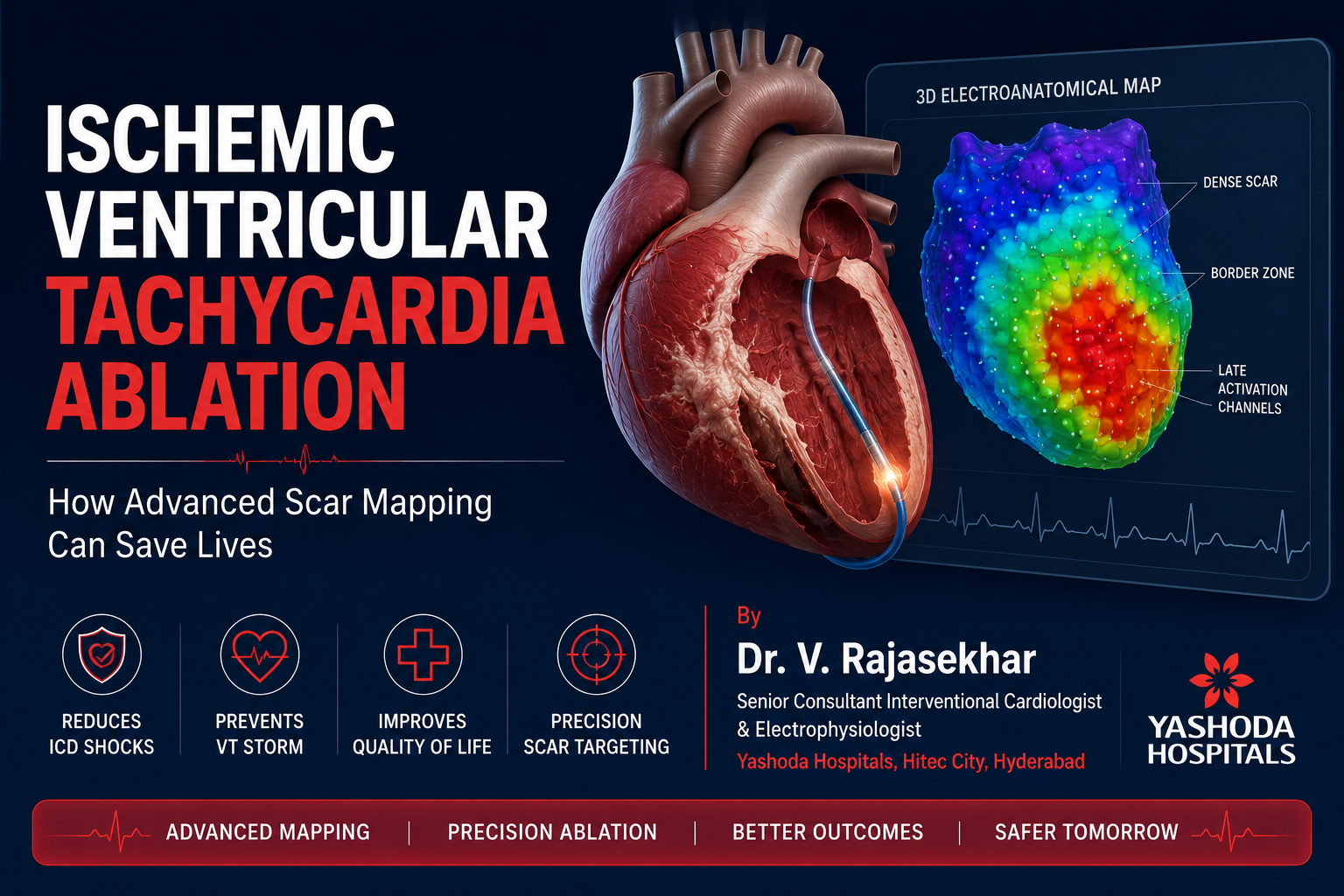
The Role of Advanced 3D Electroanatomical Mapping
Modern VT ablation depends heavily on advanced mapping technology.
Three-dimensional mapping systems create an electrical map of the heart that allows cardiologists to identify:
- Healthy myocardium
- Dense scar tissue
- Border zones
- Slow conduction channels
- Critical isthmus pathways
- Areas of late electrical activation
This information guides highly precise catheter ablation while preserving healthy heart muscle.
Scar Mapping: Finding the Hidden Problem
Voltage mapping identifies damaged heart tissue based on electrical signals.
Healthy myocardium produces strong electrical signals.
Scar tissue produces very weak or absent electrical activity.
The resulting map clearly outlines:
- Dense scar
- Border zones
- Surviving muscle channels
These surviving channels are often responsible for sustaining ventricular tachycardia.
Isochronal Late Activation Mapping (ILAM)
One of the most significant advances in VT ablation is Isochronal Late Activation Mapping (ILAM).
Rather than simply identifying scar, ILAM detects deceleration zones where electrical impulses slow dramatically.
These zones often represent:
- Critical VT isthmus
- Slow conduction pathways
- Re-entry circuits
Targeting these areas dramatically improves procedural success.
How VT Ablation Is Performed
The procedure is performed in a specialized electrophysiology laboratory.
Step 1 – Electrophysiology Study
Thin catheters are inserted through the veins and guided into the heart.
Electrical signals are recorded from multiple locations.
Step 2 – Creating a 3D Map
Thousands of electrical points are collected to create a high-resolution map of the ventricles.
The map reveals scar tissue and slow conduction zones.
Step 3 – VT Induction
Controlled stimulation attempts to reproduce the patient’s ventricular tachycardia.
This confirms the location of the abnormal circuit.
Step 4 – Radiofrequency Ablation
Heat energy is delivered through the catheter to destroy abnormal electrical tissue.
The goal is to interrupt every critical pathway responsible for VT.
Step 5 – Repeat Testing
After ablation, cardiologists again attempt to induce VT.
If no VT can be induced, the procedure is considered successful.
Benefits of VT Ablation
Successful catheter ablation can provide numerous benefits, including:
- Significant reduction in ICD shocks
- Fewer VT episodes
- Reduced hospital admissions
- Improved quality of life
- Better exercise tolerance
- Lower risk of VT storm
- Potential improvement in heart function
- Reduced dependence on antiarrhythmic medications
Is VT Ablation Safe?
Like all cardiac procedures, VT ablation carries certain risks, including bleeding, vascular injury, stroke, or damage to the heart.
However, when performed by experienced electrophysiologists using advanced mapping systems, the procedure is generally safe and highly effective.
For patients suffering recurrent VT or VT storm, the benefits often far outweigh the risks.
Recovery After VT Ablation
Most patients remain in the hospital for one to three days.
Recovery usually involves:
- Observation
- Medication adjustment
- ICD reprogramming
- Gradual return to daily activities
Regular follow-up ensures long-term rhythm stability.
Why Choose Dr. V. Rajasekhar?
Dr. V. Rajasekhar is a leading interventional cardiologist at Yashoda Hospitals, Hitec City, Hyderabad, with extensive expertise in advanced electrophysiology and complex coronary interventions.
His areas of expertise include:
- Ventricular Tachycardia Ablation
- Complex Scar Modification
- 3D Electroanatomical Mapping
- Primary Angioplasty
- Complex Coronary Angioplasty
- IVUS & OCT Guided PCI
- Left Main Interventions
- High-Risk Coronary Procedures
- Structural Heart Interventions
Using the latest mapping technologies and evidence-based treatment strategies, Dr. Rajasekhar provides comprehensive care for patients with life-threatening cardiac arrhythmias.
Final Thoughts
Ischemic Ventricular Tachycardia is one of the most dangerous consequences of a previous heart attack. While ICDs save lives during VT episodes, they do not prevent the arrhythmia from recurring.
Modern 3D electroanatomical mapping, scar modification, and catheter ablation have revolutionized treatment by identifying and eliminating the electrical circuits responsible for VT.
For patients experiencing recurrent ICD shocks or VT storm, timely evaluation by an experienced electrophysiologist can dramatically improve both survival and quality of life.
Early diagnosis, advanced mapping, and precision ablation are transforming the future of ventricular arrhythmia care—helping patients move from fear and repeated shocks to lasting rhythm control and renewed confidence.