 090003 25483
 Yashoda Hospitals Hitec City Hyderabad, Kothaguda, Telangana 500084
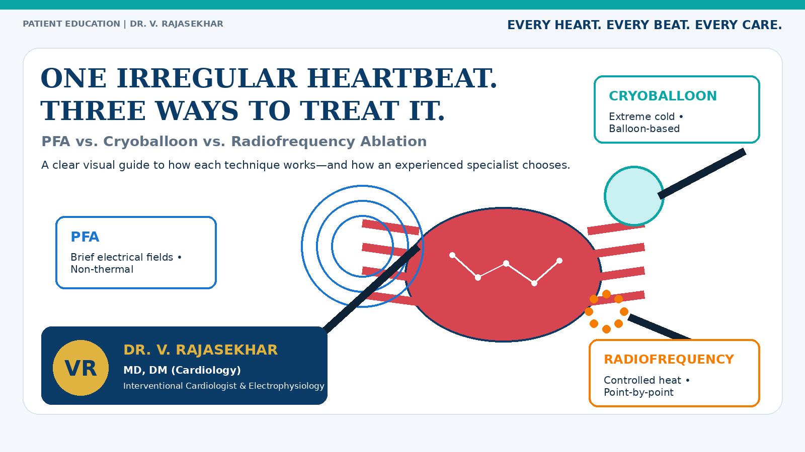
One Irregular Heartbeat. Three Ways to Treat It.
When people hear “heart ablation,” it can sound like one standard procedure. In reality, catheter ablation is a treatment strategy, not a single tool. An electrophysiology team may use pulsed electrical fields, freezing temperatures or controlled heat to interrupt the abnormal electrical pathways that keep an arrhythmia alive.
The most useful question is not “Which machine is the newest?” It is: “Which energy source, catheter design and lesion strategy best match my rhythm, my heart anatomy and the team treating me?”
Inside this guide
- What cardiac ablation is trying to achieve
- How PFA, cryoballoon and radiofrequency work
- What happens during the procedure
- The real strengths and limitations of each option
- How doctors choose the right strategy
- Safety, recovery and the blanking period
- Questions to ask before deciding
1. What Is Cardiac Ablation Actually Trying to Do?
Your heartbeat is coordinated by an electrical system. In an arrhythmia, signals may start in the wrong place, travel through an abnormal circuit or repeatedly re-enter tissue that should not keep the rhythm going. Catheter ablation creates small, controlled areas that no longer conduct the unwanted signal.
For atrial fibrillation (AF), a common target is the tissue around the pulmonary veins, where many triggering signals arise. Creating a ring of electrical isolation around these veins is called pulmonary-vein isolation (PVI). For atrial flutter, supraventricular tachycardia or some ventricular arrhythmias, the target and lesion pattern may be different.
|
KEY IDEA Ablation treats an electrical pathway—not the whole heart. Precision in mapping, catheter position and lesion verification matters as much as the energy source. |
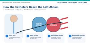
A simplified route: femoral vein → right atrium → transseptal access → left atrium.
2. Think of Ablation as Drawing a “Do Not Cross” Line
Imagine the abnormal signal as a vehicle repeatedly entering a restricted road. Ablation creates a carefully placed boundary. PFA, cryoballoon and radiofrequency can all build that boundary, but each uses a different physical language:
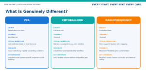
- PFA uses short electrical pulses that create irreversible electroporation in targeted cells.
- Cryoballoon ablation freezes tissue using a balloon positioned at the pulmonary-vein opening.
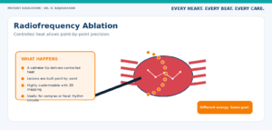
- Radiofrequency ablation heats tissue through a catheter tip and usually builds lesions point by point.
This is why a technology can be excellent without being the automatic choice. A simple circular target may suit a balloon or multi-electrode workflow. A scar-related circuit, a repeat procedure or an additional atrial tachycardia may require detailed mapping and customised lesions.
3. What Happens During an Ablation Procedure?
Most catheter ablations are performed in a cardiac electrophysiology laboratory. After anaesthesia or sedation, thin catheters are advanced through a blood vessel—commonly from the groin—into the heart. The team records electrical activity, confirms the target, delivers the chosen energy and tests whether the abnormal pathway has been interrupted.
|
1 Access A small sheath is placed in a vein, usually in the groin. |
2 Mapping Electrical recordings and imaging identify the treatment target. |
3 Ablation PFA, cryo or radiofrequency energy is delivered according to the strategy. |
4 Verification The team checks isolation or interruption of the rhythm circuit. |
4. Pulsed Field Ablation (PFA): A Non-Thermal Approach
How it works
PFA delivers very brief, high-voltage electrical pulses. These pulses create microscopic pores in cell membranes—a process called electroporation. When designed to be irreversible, the targeted heart-muscle cells lose their ability to conduct the unwanted signal and form the intended lesion.
Why patients and doctors are interested
- Energy delivery can be rapid in suitable pulmonary-vein isolation workflows.
- The mechanism does not primarily depend on deliberate heating or freezing.
- Its relative tissue selectivity changes the discussion about injury to nearby structures.
- Evidence and real-world experience are expanding quickly.
Where the limits still matter
- Most mature experience is in atrial fibrillation and pulmonary-vein isolation.
- PFA systems are not identical; catheter shape, pulse sequence and mapping integration matter.
- The procedure still carries risks related to vascular access, transseptal puncture, clot formation and recurrence.
- Long-term durability and best-use strategies continue to evolve.
|
HONEST TAKE PFA is promising, not magical. The outcome still depends on patient selection, complete electrical isolation, lesion durability and team experience. |
5. Cryoballoon Ablation: Freezing a Circular Barrier
How it works
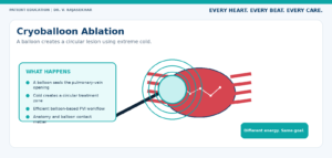
A balloon catheter is positioned at the opening of a pulmonary vein and cooled to create a circumferential lesion. In suitable anatomy, this can isolate the vein without building the entire circle point by point.
Why it remains an established option
- A well-established option for pulmonary-vein isolation in selected AF patients.
- A reproducible balloon-based workflow in experienced hands.
- Efficient when the balloon can achieve good contact and occlusion.
- Supported by extensive clinical use over many years.
Where it may be less flexible
- Designed mainly for circular pulmonary-vein targets.
- Pulmonary-vein anatomy can influence balloon contact.
- Phrenic-nerve monitoring is important near the right-sided veins.
- Thermal safety precautions remain essential.
6. Radiofrequency Ablation: Point-by-Point Precision
How it works
Radiofrequency ablation delivers controlled heat through the tip of a catheter. The operator creates lesions point by point, often using contact-force feedback, power parameters and three-dimensional mapping to guide placement.
Why RF remains central to electrophysiology
- Highly versatile for many rhythm disorders beyond pulmonary-vein isolation.
- Allows customised lines and focal lesions for complex circuits.
- Useful for detailed mapping, targeted touch-up and treatment beyond a balloon-shaped target.
- Supported by decades of experience and continuous technology improvement.
Important trade-offs
- Point-by-point treatment can require more mapping and lesion-delivery time.
- Thermal injury to nearby structures must be actively prevented.
- Durability depends on lesion continuity, depth, contact and catheter stability.
- Outcomes depend on the rhythm type, heart substrate and operator experience—not simply the power setting.
7. Side-by-Side Comparison

|
BOTTOM LINE The catheter and strategy are a package. Mapping quality, lesion verification, anaesthesia, anticoagulation management, team experience and follow-up all influence safety and effectiveness. |
8. How Doctors Decide: A Clinical Compass, Not a Popularity Contest
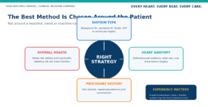
A heart-rhythm specialist may consider:
- The exact rhythm and whether the main target is pulmonary-vein isolation or a more complex circuit.
- Whether this is a first ablation or a repeat procedure.
- Atrial size, pulmonary-vein anatomy, scar and previous heart surgery.
- Heart function, kidney or lung disease, bleeding risk and other medical conditions.
- Whether detailed mapping, additional lines, focal lesions or a combined strategy will be required.
- The technologies the centre uses regularly and the team’s safety systems.
|
EXPERIENCE MATTERS An experienced team using an established method may be safer and more effective than choosing a newer technology that is not yet embedded in the centre’s routine workflow. |
9. Three Patient Scenarios That Show Why the Answer Can Differ
|
Scenario A: First-time intermittent AF A patient has symptomatic paroxysmal AF, suitable pulmonary-vein anatomy and no previous ablation. The discussion may reasonably include PFA, cryoballoon or RF, depending on the centre’s systems and experience. |
|
Scenario B: Recurrent AF after a previous procedure Mapping shows a reconnected vein and an additional atrial tachycardia. A point-by-point RF strategy—or a combined approach—may provide greater flexibility for touch-up and treatment of the additional circuit. |
|
Scenario C: A rhythm that is not AF A patient has a focal supraventricular tachycardia or typical atrial flutter. The target is not a circular pulmonary-vein lesion, so a precise focal or line-based strategy may be more suitable. |
|
EDUCATIONAL EXAMPLES These fictional scenarios explain clinical reasoning. Real treatment requires review of ECGs, monitoring, imaging, medication history and individual risks. |
10. Safety: What Patients Should Hear Clearly
All catheter ablations are invasive procedures. Serious complications are uncommon in experienced centres, but “minimally invasive” does not mean “zero risk.” The exact risk depends on the arrhythmia, procedure type, patient health and hospital experience.
- Bleeding, bruising or blood-vessel injury at the access site
- Pericardial effusion or cardiac tamponade
- Stroke or transient ischaemic attack
- Anaesthesia-related complications
- Damage to nearby structures, depending on the target and energy source
- Pulmonary-vein narrowing, phrenic-nerve injury or oesophageal injury in specific thermal contexts
- Need for repeat treatment if the rhythm returns or lesions reconnect
|
ASK YOUR SPECIALIST What is this centre’s complication rate for patients with a rhythm and procedure similar to mine? |
11. Recovery and the “Blanking Period”
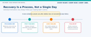
Many patients are surprised to experience extra beats, brief palpitations or short rhythm episodes after ablation. During the early healing period, inflammation can temporarily irritate the heart. An episode during this time does not automatically mean the procedure has failed.
Medication decisions are individual. Anticoagulation is guided by stroke risk and the treating physician’s plan—not only by how normal the rhythm feels. Never stop a blood thinner or rhythm medicine without explicit medical advice.
12. Questions to Take to Your Consultation
- What exact arrhythmia am I being treated for?
- Is the main target pulmonary-vein isolation, or will I need additional lesions?
- Why are you recommending PFA, cryoballoon or RF for my anatomy and rhythm?
- How experienced is the team with this specific system?
- What are the important success, repeat-procedure and complication considerations in my case?
- Will I need general anaesthesia or deep sedation?
- How long should I continue anticoagulation and rhythm medication?
- What symptoms are expected after discharge, and which require urgent help?
- How will rhythm recurrence be monitored?
- What is the plan if the first procedure does not fully control the arrhythmia?
13. Frequently Asked Questions
Is PFA safer than cryo or RF?
PFA’s non-thermal mechanism may reduce certain collateral-injury concerns. However, it still carries procedural risks, and safety depends on the patient, catheter system, lesion plan and clinical team.
Which ablation has the highest success rate?
There is no universal winner. The type of AF, atrial disease, previous treatment, monitoring method and follow-up duration strongly affect the comparison.
Is the newest method always the best method?
No. A mature technology used by a highly experienced team may be the strongest choice in one case. A newer technology may offer an advantage in another.
Can atrial fibrillation return after ablation?
Yes. Veins can reconnect, new triggers may appear or underlying atrial disease may progress. Some patients need medication, cardioversion or repeat ablation.
Will I stop blood thinners after ablation?
Not automatically. Stroke-prevention decisions remain based on individual risk and the treating physician’s plan.
How soon can I return to work?
Many people return to light activity within several days, but the timing depends on access-site healing, job demands and individual instructions.
Does ablation cure the underlying tendency to AF?
Ablation can reduce rhythm episodes, but weight, blood pressure, sleep apnoea, alcohol, diabetes and fitness can still influence recurrence.
Who should perform the procedure?
A trained electrophysiologist supported by an experienced hospital team with appropriate mapping, anaesthesia, anticoagulation, imaging and emergency systems.
14. The Final Takeaway
PFA, cryoballoon and radiofrequency ablation are not competing answers to one identical question. PFA brings a non-thermal mechanism and rapidly expanding experience. Cryoballoon offers an established balloon-based route to pulmonary-vein isolation. Radiofrequency provides customised, point-by-point precision across a wide range of rhythm problems.
The best decision is the one that makes clinical sense for your arrhythmia, anatomy, overall health and treating team. A good consultation should leave you understanding not only what your doctor recommends—but why.
About Dr. V. Rajasekhar
|
Senior Interventional Cardiologist & Electrophysiology Specialist Yashoda Hospitals, Hitec City, Hyderabad His clinical work includes cardiac-rhythm evaluation, electrophysiology studies, catheter ablation, complex coronary intervention, image-guided PCI and cardiac-device implantation. His consultation approach focuses on identifying the exact rhythm problem and explaining why a particular treatment strategy fits the individual patient. Doctor-led focus: personalised assessment rather than technology-led promotion. |
|
NEED A PERSONALISED RHYTHM EVALUATION? Patients experiencing repeated palpitations, an irregular heartbeat, unexplained breathlessness, dizziness, fatigue or recurrent atrial fibrillation can seek an individual assessment with Dr. V. Rajasekhar at Yashoda Hospitals, Hitec City, Hyderabad. |
Medical disclaimer: This article is intended for general patient education and does not replace an individual consultation, diagnosis or treatment recommendation. The suitability, benefits and risks of cardiac ablation must be assessed by a qualified cardiologist or electrophysiologist after reviewing the patient’s complete medical condition.