Introduction: A Case That Was Anything but Routine
In cardiology, we often prepare for the expected — but every now and then, a case reminds us just how unpredictable the human body can be.
A 63-year-old gentleman walked into our OPD with complaints that may sound ordinary: breathlessness on even minimal exertion and a sense of giddiness for the past week. But what unfolded was a rare anatomical twist that turned a seemingly straightforward pacemaker implantation into a challenging — and deeply rewarding — procedure.
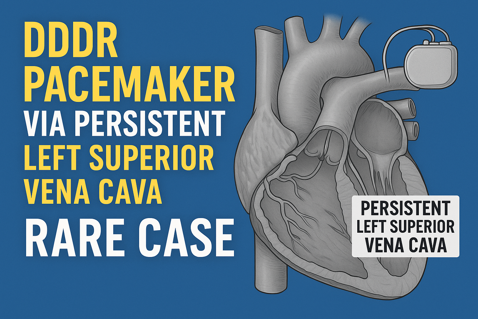
Let’s take you through the journey of how we implanted a dual-chamber rate-responsive pacemaker (DDDR) via a Persistent Left Superior Vena Cava (PLSVC) — an anomaly seen in less than 0.5% of the population.
The First Signs: A Heart Out of Sync
When we first saw him, he was stable but noticeably bradycardic. His heart rate hovered around 38 beats per minute. For someone who wasn’t on any medication that slows down the heart, this was a red flag.
Key symptoms:
-
Shortness of breath even while doing everyday activities
-
A feeling of lightheadedness, almost like he was going to faint
Known history:
-
Hypertension (HTN)
-
Type 2 Diabetes Mellitus (T2DM)
We performed an ECG immediately — and there it was: Complete Heart Block. The atria and ventricles were no longer communicating properly, leaving the heart to rely on a dangerously slow escape rhythm.
Further Investigations: What’s Going on Inside?
An echocardiogram showed something reassuring: the heart muscle was pumping well. There were no regional wall motion abnormalities (RWMA), and the left ventricular ejection fraction was normal.
All routine labs were within range, and the patient was otherwise healthy. This meant one thing — we had a strong candidate for permanent pacemaker implantation.
We geared up for what we expected to be a fairly routine DDDR pacemaker procedure. But during the procedure, we hit an unexpected detour.
The Twist: A Guidewire That Went the Wrong Way
As we accessed the left subclavian vein and introduced the guidewire, something didn’t feel right.
Instead of heading toward the right atrium, the wire took a leftward and downward course, looping back and entering the heart from an unusual angle.
That’s when the suspicion hit — this wasn’t our standard superior vena cava. We were likely dealing with a Persistent Left Superior Vena Cava (PLSVC).
A venogram confirmed it: the patient’s left upper body venous drainage wasn’t following the usual route. Instead, it drained into the coronary sinus and then into the right atrium — a rare congenital anomaly that had gone unnoticed his entire life.
What Is a Persistent Left Superior Vena Cava?
In the vast majority of people, the left superior vena cava disappears during fetal development. But in a tiny percentage, it remains — silently. Most people with PLSVC never know they have it unless they undergo procedures like central line insertions or pacemaker implantations.
In this case, PLSVC meant a longer, more tortuous route for our pacemaker leads, and the coronary sinus was notably dilated — both of which can make lead positioning tricky.
But the team was ready.
The Game Plan: Adjusting for the Anatomy
After discussing the situation with the patient and his family, we went ahead with a DDDR pacemaker — a dual-chamber device that can pace both the atria and ventricles while adjusting the rate based on physical activity.
Here’s how we tackled it step-by-step:
✅ 1. Navigating the Route
Using specially curved stylets and careful fluoroscopic guidance, we advanced the leads:
-
Through the PLSVC
-
Into the coronary sinus
-
Then into the right atrium
-
And finally, into the right ventricle
✅ 2. Securing the Leads
-
The ventricular lead was actively fixed in the right ventricular apex
-
The atrial lead was positioned in the right atrial appendage
Despite the challenging route, the lead thresholds and impedance were within ideal limits. There was no phrenic nerve stimulation — a known risk when navigating through the coronary sinus.
✅ 3. Final Steps
The generator was secured in the left pectoral pocket, and post-procedure testing confirmed appropriate AV synchrony and stable pacing.
Recovery & Follow-up
The patient recovered smoothly and was discharged within 48 hours. Post-implant interrogation showed:
-
Excellent sensing and pacing thresholds
-
Synchronous AV pacing
-
No complications
He was advised regular follow-up, especially within the first few weeks, to monitor lead stability and device function.
What Makes This Case Special?
This wasn’t just about managing complete heart block — it was about adapting to an unexpected anatomical challenge and delivering the best outcome regardless.
Here’s why this case stands out:
-
PLSVC is rare: Less than 0.5% of the population has it.
-
It complicates pacemaker implantation: Leads must take a longer, curved path, increasing risks.
-
Pre-procedure imaging didn’t show it: It was discovered during the actual procedure, demanding on-the-spot adjustments.
But with experience, teamwork, and proper planning, we successfully completed the implantation — and gave the patient a new rhythm to life.
Takeaways for Clinicians & Learners
-
Always watch the guidewire: An unusual path could be a sign of anatomical variation like PLSVC.
-
Keep PLSVC in mind when leads fail to follow the typical course, especially on the left side.
-
Venography or intraoperative imaging is key when you suspect anomalies.
-
Don’t rush lead placement: Take the time to shape the stylet, navigate carefully, and ensure secure fixation.
-
Adaptability is everything: Knowing your anatomy is important, but being ready for what you don’t expect is even more crucial.
❓ FAQs: DDDR Pacemaker via PLSVC – What You Need to Know
1. What is a DDDR pacemaker and how is it different from a regular pacemaker?
A DDDR pacemaker is a dual-chamber, rate-responsive device. It paces both the atria and ventricles and adjusts the heart rate based on your physical activity. It offers more physiological pacing compared to single-chamber devices, which only stimulate one chamber of the heart.
2. What is Persistent Left Superior Vena Cava (PLSVC)?
PLSVC is a rare congenital venous anomaly where the vein that normally regresses in fetal life remains and drains blood from the upper body into the heart through an unusual path — typically via the coronary sinus. It’s usually harmless but can complicate procedures like pacemaker insertions.
3. How common is PLSVC?
PLSVC occurs in about 0.3% to 0.5% of the general population. Many people live their entire lives without knowing they have it — unless they undergo imaging or certain cardiac procedures.
4. Why does PLSVC make pacemaker implantation difficult?
The abnormal venous route creates a longer, curved path for pacemaker leads. This makes it technically challenging to position and secure the leads in the right chambers of the heart. Special tools and careful technique are required to avoid lead dislodgement or phrenic nerve stimulation.
5. How is PLSVC diagnosed during a pacemaker procedure?
It’s often discovered accidentally during the procedure when the guidewire or lead follows an unexpected route. In such cases, contrast venography or intraoperative fluoroscopy is used to confirm the diagnosis.
6. Is pacemaker placement via PLSVC safe?
Yes, when performed by experienced cardiologists using proper tools and techniques, DDDR pacemaker implantation via PLSVC is safe and effective. Most patients recover well and enjoy normal pacing function.
7. Will having PLSVC affect the performance of the pacemaker?
Not at all. Once the leads are placed correctly and the device is programmed properly, the pacemaker works just like it would in someone without PLSVC.
8. Are there any long-term complications with PLSVC and pacemakers?
Long-term outcomes are generally good. However, close follow-up is recommended in the early weeks to monitor for:
-
Lead dislodgement
-
Sensing or pacing issues
-
Device migration
9. Can PLSVC be detected before the pacemaker procedure?
Sometimes. If a patient has had prior imaging (like CT or MRI of the chest), PLSVC might be identified beforehand. However, it’s often missed until procedures like pacemaker implantation reveal it.
10. How long does it take to recover from pacemaker implantation via PLSVC?
Most patients are discharged within 24–48 hours and return to normal activity in a few days, with some restrictions. Full recovery usually takes 1–2 weeks, and follow-up checks are essential to ensure everything is functioning as expected.