 090003 25483
 Yashoda Hospitals Hitec City Hyderabad, Kothaguda, Telangana 500084

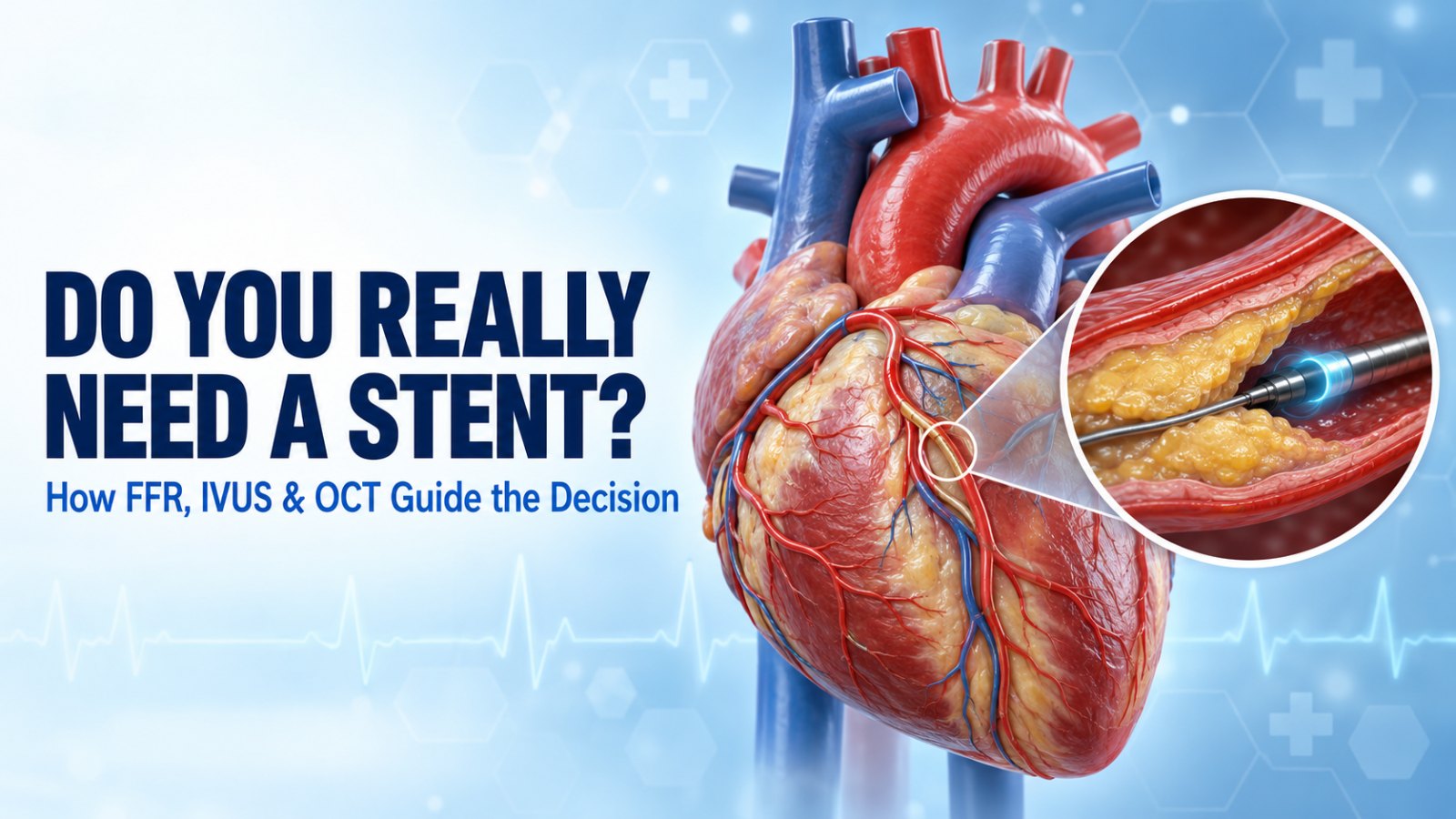
Doctor, you found a blockage. Does that mean I need a stent? It is one of the most common questions in any cardiology clinic, and one of the most important. The honest answer is: not always. Some narrowings that look worrying on an angiogram are not actually starving the heart of blood, and some that look modest are far more significant than they appear.
Modern cardiology has tools that let us stop guessing. FFR measures whether a blockage is truly limiting blood flow. IVUS and OCT let us see the artery from the inside, so that when a stent is needed, it is the right size and placed in the right spot. This article explains how these tests work, in plain language, so you can walk into your consultation with the right questions.
In this article
Why an Angiogram Alone Can Mislead
A coronary angiogram is an X-ray movie of dye flowing through your heart arteries. It is excellent at showing where narrowings are. But it has two built-in limitations:
- It is a shadow, not a photograph. The angiogram shows the outline of the dye-filled channel (doctors call it a “lumenogram”). It does not show the artery wall or the plaque itself.
- Percentages are estimates. “70% blockage” is usually a visual estimate. Two experienced cardiologists can look at the same picture and differ by 10–20%. A narrowing that looks severe from one angle can look mild from another.
Most importantly, the angiogram does not tell us whether the narrowing is actually reducing blood supply to the heart muscle. That is the question that decides whether a stent will help you.
Key takeaways
- A blockage percentage on an angiogram is an estimate, not a verdict.
- FFR measures blood flow across a blockage; a stent helps mainly when flow is significantly reduced.
- IVUS and OCT show the artery wall from inside, helping choose the right stent size and confirm a good result.
- Large clinical trials show these tools can mean fewer unnecessary stents and fewer repeat problems.
FFR: Measuring Whether a Blockage Really Matters
Fractional Flow Reserve (FFR) is a pressure measurement taken during the angiogram. A guidewire, thinner than a pencil lead, with a tiny pressure sensor at its tip is passed beyond the narrowing. It compares the pressure after the blockage with the pressure before it.
- An FFR of 1.0 means no pressure drop at all.
- An FFR of 0.80 or below generally means the blockage is significantly limiting blood flow, and treating it is likely to help.
- An FFR above 0.80 usually means the narrowing can be safely managed with medicines and lifestyle changes, without a stent.
A related measurement called iFR (instantaneous wave-free ratio) works on the same principle but does not need the medicine (adenosine) that FFR uses to maximise blood flow. A value of 0.89 or below is considered significant.
What the evidence says
In the landmark FAME trial (New England Journal of Medicine, 2009), patients with multiple blockages were treated either based on how the angiogram looked or based on FFR. The FFR-guided group received fewer stents and had a lower rate of death, heart attack or repeat procedures at one year. In simple terms: measuring before stenting led to fewer stents and better results.
IVUS and OCT: Seeing the Artery From the Inside
Once a decision to stent has been made, a new question arises: how exactly should it be done? This is where intravascular imaging comes in.
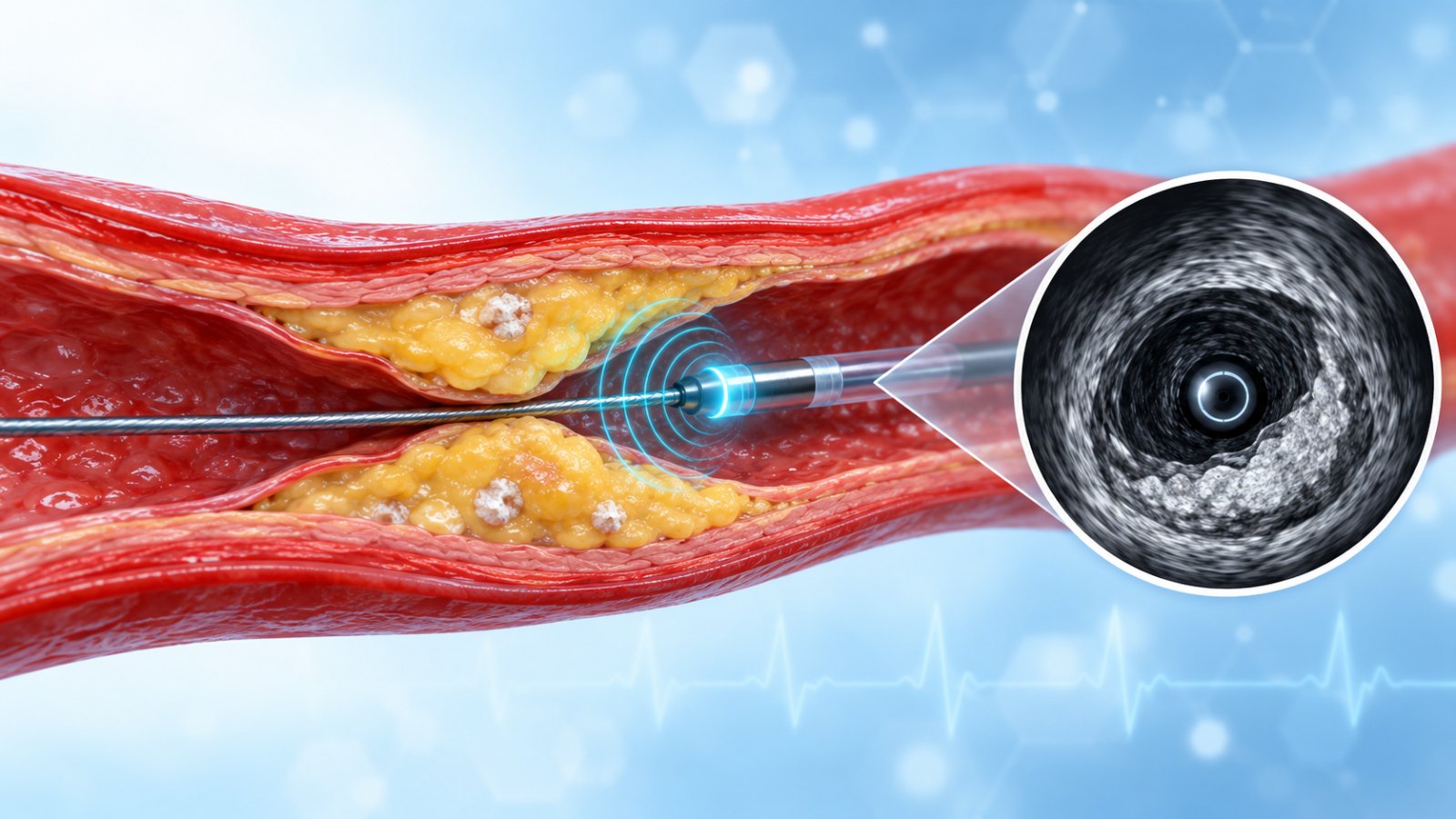
IVUS (Intravascular Ultrasound)
A miniature ultrasound probe on a catheter. It sees deep into the artery wall, measures the true vessel size, and shows how much plaque and calcium is present. Very useful in long blockages and left main disease.
OCT (Optical Coherence Tomography)
Uses near-infrared light for extremely sharp, high-resolution images, about ten times finer than IVUS. Excellent for checking how well a stent has expanded and whether its edges are sitting perfectly.
Why this matters to you as a patient
A stent that is too small, not fully expanded, or that stops short of the diseased segment is more likely to narrow again or form a clot later. Imaging helps the cardiologist:
- Choose the correct stent diameter and length, measured rather than guessed.
- Identify heavy calcium that needs preparation first (for example with rotablation, laser or intravascular lithotripsy).
- Confirm the stent is fully expanded and well apposed to the artery wall before you leave the cath lab.
The RENOVATE-COMPLEX-PCI trial (NEJM, 2023) found that in complex blockages, imaging-guided stenting reduced the risk of future target-vessel problems by roughly a third compared with angiography-guided stenting. International guidelines from the European Society of Cardiology now recommend intravascular imaging for complex procedures such as left main and bifurcation stenting.
FFR vs IVUS vs OCT at a Glance
| Tool | Main question it answers | How it works | Typical use |
|---|---|---|---|
| FFR / iFR | Does this blockage need treatment? | Pressure wire across the narrowing | Borderline (40–90%) blockages, multiple blockages |
| IVUS | How big is the artery and what is the plaque like? | Ultrasound from inside the artery | Left main, long lesions, calcium, stent sizing |
| OCT | Is the stent perfectly expanded and positioned? | High-resolution light-based imaging | Stent optimisation, bifurcations, stent failure |
Common Myths About Stents, Busted
“Every blockage above 70% needs a stent.”
Not necessarily. Symptoms, FFR results and the overall picture matter more than a single number. In stable patients, many blockages are best treated with medicines alone.
“If they’re testing with FFR, they’re trying to sell me a stent.”
It is usually the opposite. FFR exists precisely to avoid stents that will not help. It protects patients from overtreatment.
“Imaging just adds cost without benefit.”
In complex cases, imaging lowers the chance of the stent failing later, which can save you a repeat procedure, a hospital stay, or worse.
“Heart attacks always need FFR first.”
No. During an active heart attack, the blocked artery is opened immediately. Physiology testing is mainly for stable patients and for non-culprit arteries.
Questions to Ask Your Cardiologist
- Is my blockage causing my symptoms, or is it an incidental finding?
- Would an FFR or iFR measurement help decide whether I need a stent?
- If I need a stent, will IVUS or OCT be used to size and check it?
- Is there calcium that needs special preparation before stenting?
- What are my options if I choose medicines first?
How Dr. V. Rajasekhar Approaches the Decision
Dr. V. Rajasekhar is a senior interventional cardiologist at Yashoda Hospitals, Hitec City, with 27 years of practice and is a proctor (trainer) for image-guided PCI and rotablation. His approach is simple: treat the patient, not the picture. When the need for a stent is uncertain, physiology testing guides the decision. When a stent is needed, imaging guides how it is done, especially in calcified, long, left main and bifurcation blockages.
Frequently Asked Questions
Is FFR testing painful or risky?
FFR is done through the same wrist or groin access as the angiogram. Some patients feel brief chest warmth or breathlessness from the adenosine medicine, which settles within a minute. Serious complications are rare and similar to a routine angioplasty procedure.
Can FFR be done on the same day as my angiogram?
Yes. In most cases it is done during the same sitting, so the decision to stent or not can be made right away.
What FFR value means I need a stent?
An FFR of 0.80 or below (or iFR of 0.89 or below) usually indicates a flow-limiting blockage. Your cardiologist will interpret it together with your symptoms and other findings.
What is the difference between IVUS and OCT?
IVUS uses ultrasound and sees deeper into the artery wall, which helps with vessel sizing. OCT uses light and gives much sharper images, which helps check stent expansion and edges. Both improve the quality of stenting in complex cases.
Does insurance cover FFR, IVUS or OCT?
Coverage varies between insurers and policies. Our team can help you check with your insurer before the procedure.
If I don’t get a stent, will my blockage get worse?
Plaque can progress, which is why medicines, cholesterol control, blood pressure and sugar control, quitting tobacco and regular follow-up are essential. Many patients remain stable for years with good medical therapy.
Not sure if you need a stent?
Get a clear, evidence-based opinion from Dr. V. Rajasekhar at Yashoda Hospitals, Hitec City. Bring your angiogram CD and reports.
Call 090003 25483Book a ConsultationYashoda Hospitals, Hitec City, Kothaguda, Hyderabad 500084
This article is for general education and does not replace a personal consultation. Treatment decisions depend on your individual symptoms, test results and overall health.